Designed to Perform
The hand is one of the most complex and most frequently utilized parts of the human musculoskeletal system. It is composed of a multitude of closely balanced structures, e.g. bones, muscles, ligaments, tendons, joints, and nerves. The goal of hand surgery is to minimize the impact of hand diseases and injuries on daily activities and to restore and obtain as much function as possible.
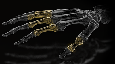
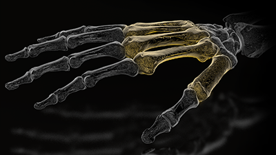
TriLock 1.5
One of the smallest polyaxial locking systems in the hand.
Unexpected Solutions
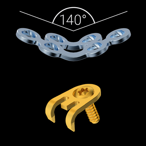
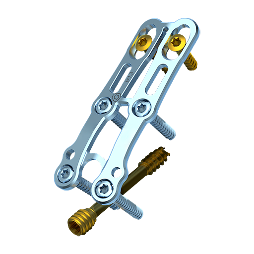
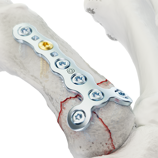
Scaphoid Plate 1.5 & Hookplate.
No fracture untreated
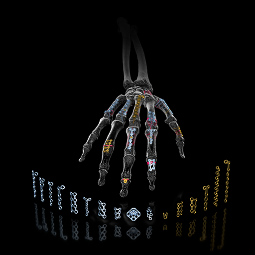
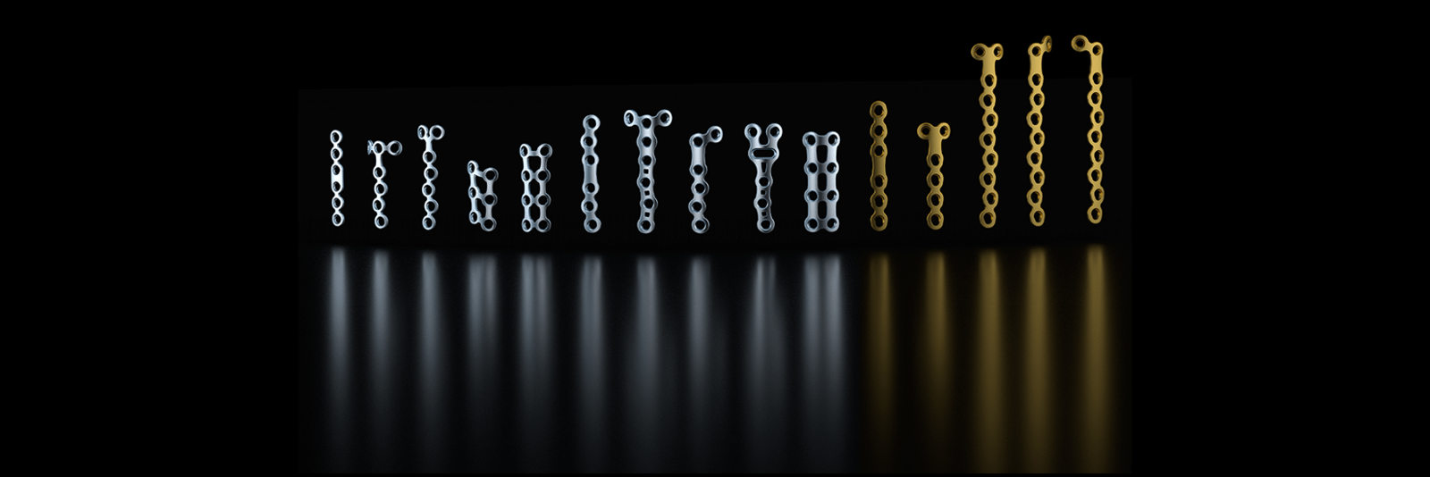
The complete Hand System 1.2 - 2.3 provides tools to face any fracture, regardless of complexity.
TriLock
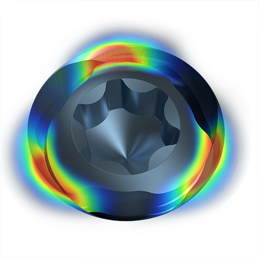
Patented TriLock locking technology provides multidirectional locking of the screw in the plate.
Treatment Concept
Over the past decades, locking plates have established themselves in the treatment of fractures in the hand and wrist. The increasing number of hand fracture systems that include locking plates reflect the trend of hand surgeons to use internal fixation systems, especially for complex fractures. The treatment with angular stable systems has the advantage of achieving a more stable fixation in fractures with bone loss and of having a reduced risk of secondary displacement. Additionally, they offer the possibility of indirect reduction of metaphyseal fractures. Due to the complex anatomy of the hand in general and the phalanges in particular, the plates should be as small as possible to spare soft tissue, but strong enough to bridge a defect zone. The aim of an osteosynthesis on the hand is to achieve an exercise-stable fracture treatment. This enables early mobilization of the hand.

TriLock T-Plate, 5/4 Hole 1.2/1.5
Design Highlights
-
Internal fixator principle for early mobilization
-
Double bars between screw holes for increased torsional stability
-
Double row T-plate: more screw options for support in the subchondral area
-
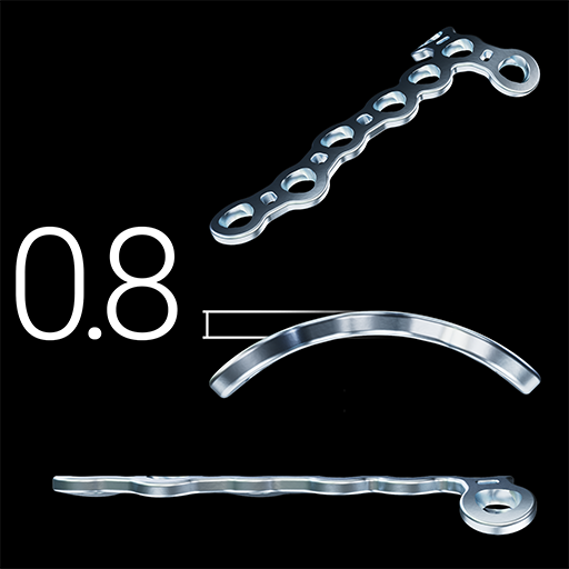
Low plate profile with 0.8 mm thickness
Features
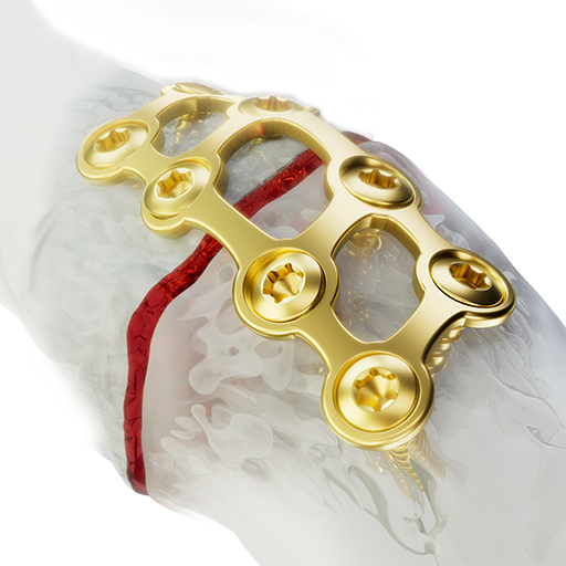
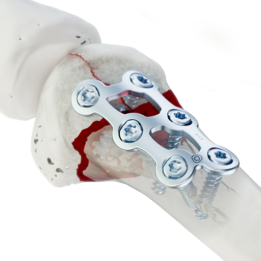
TriLock Scaphoid Plate 1.2/1.5
Highlights
-
High degree of stability due to grid structure and up to three TriLock screws on each side of the non-union
-
Anatomically preshaped to correct humpback deformity
-
Two middle bars to keep the bone graft in place
-
Completion of the scaphoid portfolio for non-compressible fractures e.g. humpback deformities, non- unions etc.
Features
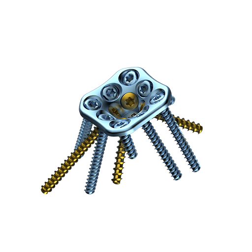
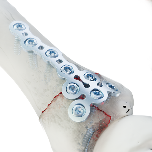
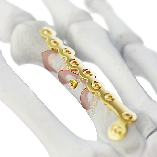
Compression Hook Plate 1.2/1.5
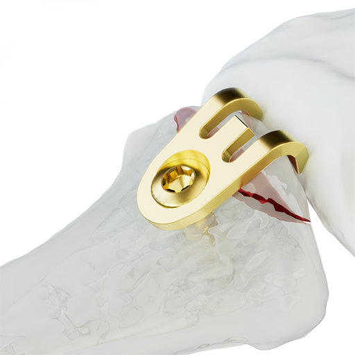
Design Highlights
-
Improved anatomical fit.
-
First distal screw row for support of the central aspect of the radio carpal joint.
-
Second distal screw row provides stabilization of the dorsal rim.
Features
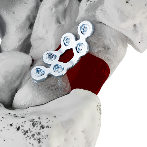
Grid Plates 1.2/1.5
Design Highlights
-
Improved anatomical fit.
-
First distal screw row for support of the central aspect of the radio carpal joint.
-
Second distal screw row provides stabilization of the dorsal rim.
Features

Condylar Plates 1.2/1.5
Design Highlights
-
Improved anatomical fit
-
First distal screw row for support of the central aspect of the radio carpal joint.
-
Second distal screw row provides stabilization of the dorsal rim.
Features
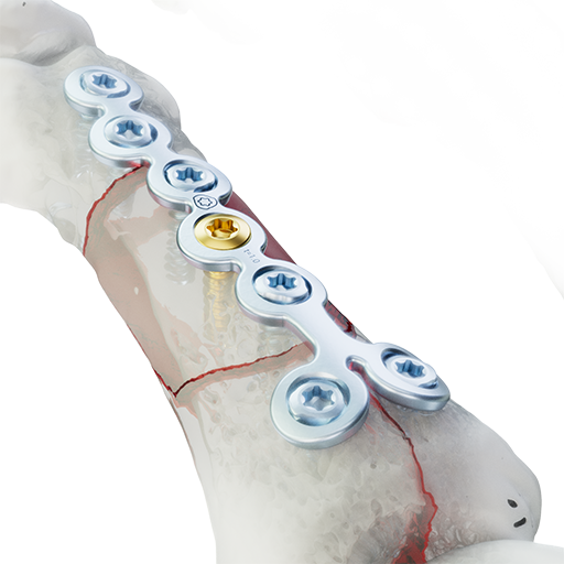
TriLock Rotation Correction Plate 2.0/2.3
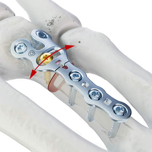
Hightlights
-
Transversal oblong hole allows up to ± 25° of rotational correction and is close to the joint to perform the osteotomy near the metaphyseal area
-
Separate screw holes (“frog design") simplify contouring in the periarticular area
Features
TriLock Y-Plate, 2.0/2.3
Design Highlights
-
Improved anatomical fit.
-
Radiolucent drill guide block available for rapid and easy angulation of screws.
-
Stabilization of the sigmoid notch and lunate facet.
MC Compression Plate 2.0/2.3
Highlights
-
Several design options: straight, L-/Y-shape
-
Low plate profile with 1.3 mm thickness
-
Broader bars in bridging zones
Features
Grid Plates 2.0/2.3
Design Highlights
-
Fractures in metacarpals and proximal phalanges
-
Comminuted fractures
-
Simple and complex intraarticular and extraarticular fractures
Features
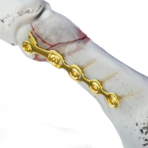
TriLock T-Plates 2.0/2.3
Design Highlights
-
Reduction of articular fracture fragments and fractures close to the joint
-
Broader bars in bridging zones
-
Low plate profile with either 1.0 or 1.3 mm thickness: choice of construct stability
Features
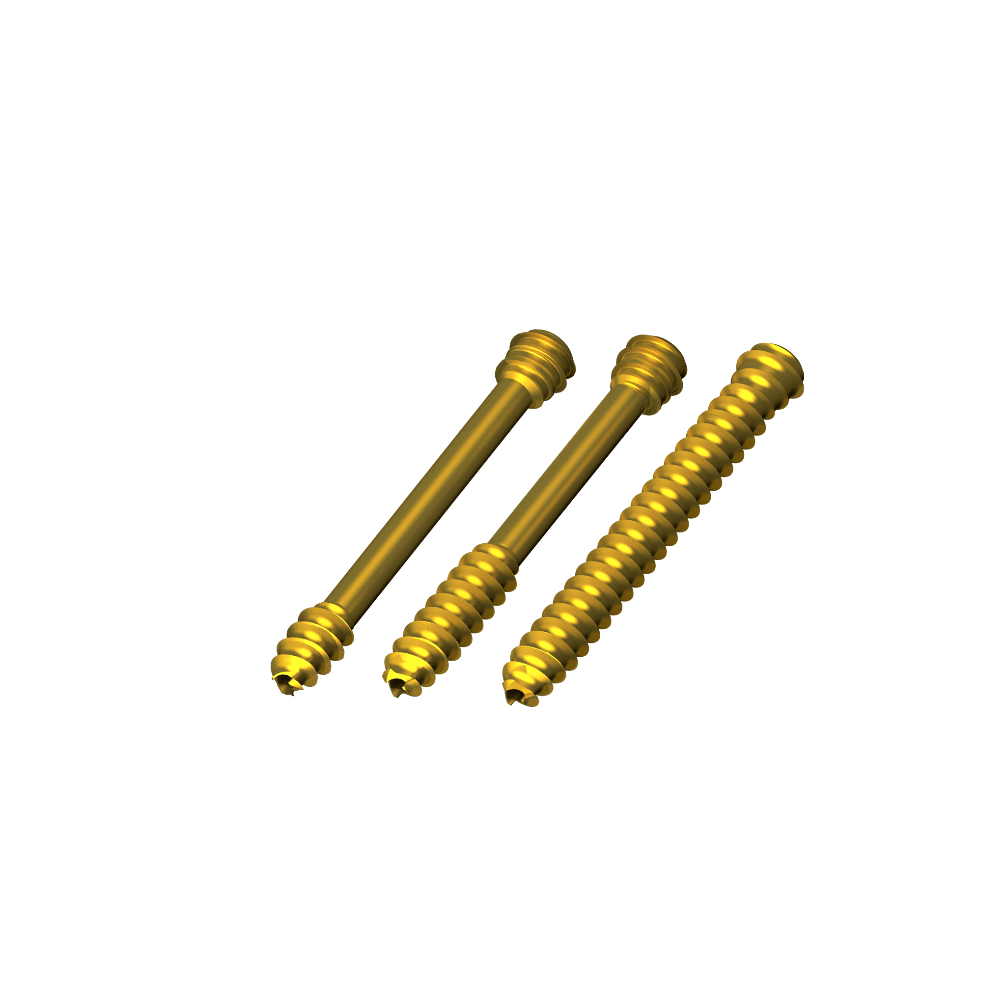
Screws

Non-locking
1.2 Cortical Screws,
HexaDrive 4

Non-locking
1.5 Cortical Screws,
HexaDrive 4

Non-locking
2.0 Cortical Screws,
HexaDrive 6

Non-locking
2.3 Cortical Screws,
HexaDrive 6

Locking
1.5 TriLock Screws,
HexaDrive 4

Locking
2.0 TriLock Screws,
HexaDrive 6
Designed to Simplify
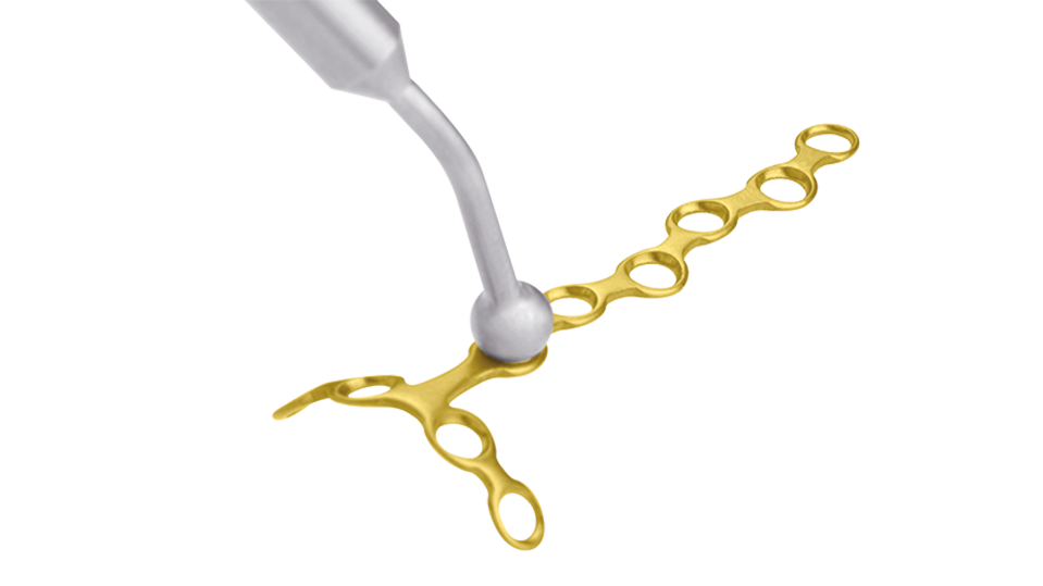
Holding and Positioning Instrument
The ball tip end of the 1.2/1.5 plate holding and positioning instrument (A-2350) facilitates positioning, moving and holding the implant on the bone and can be used with all system sizes.
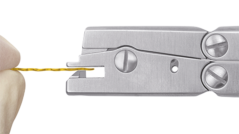
Plate Cutting
Ensure that there are no remaining plate segments in the cutting pliers (visual check). Insert the plate from the front into the open cutting pliers. Always ensure that the labeled side of the plate is facing upwards. Hold the implantable plate segment with your hand during and after cutting.
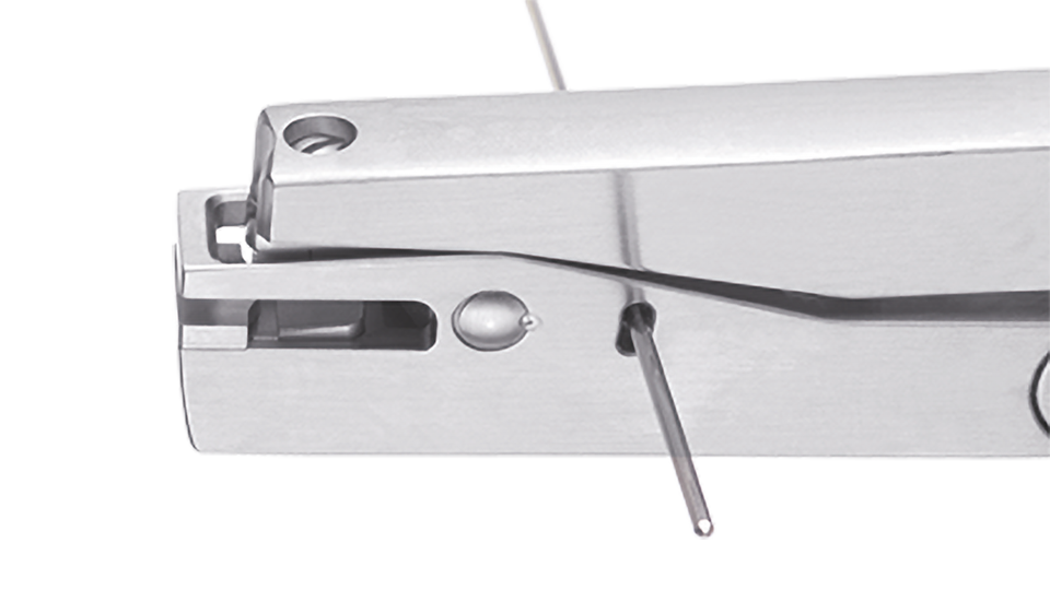
K-Wire Cutting
Shorten the K-wires by inserting the wire through the opening located on the side of the plate cutting pliers. Cut the wire by pressing the pliers.
Technological Milestones in Osteosynthesis
TriLock
Multidirectional and angular stable locking, screws can pivot freely by ± 15° in all directions
TriLockPLUS
TriLockPLUS screw holes offer the advantage of locking and compression in one step
HexaDrive
Simplified screw pick-up due to self-holding system
Designed to Organize
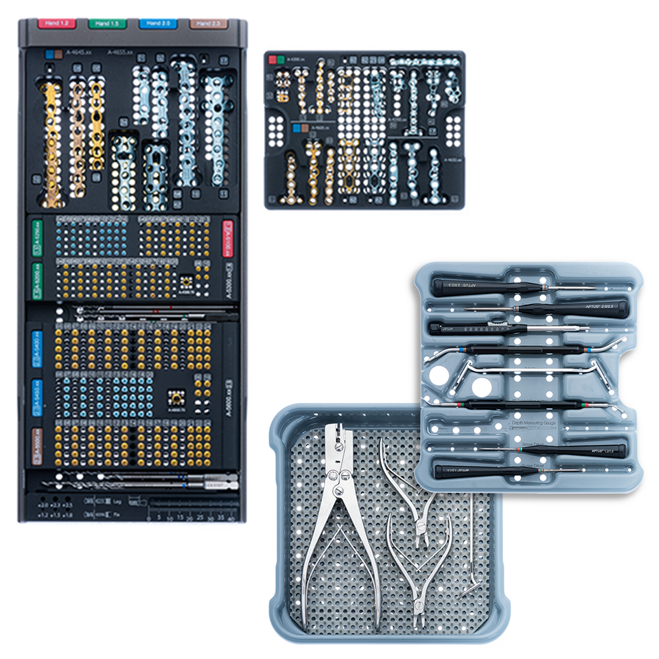
- Compact system
- Easy to use
- Lightweight components
- Validated cleaning and sterilization of the implants
Documentation
Results
- Case Study - Fixation of Mallet Fracture with APTUS 1.2 / 1.5 Hook Plate - Dr. Helen Abel 27.03.2024 1 MB Case Study Upper Extremities English
- Case Study - Scaphoid Plate: Athlete's Scaphoid Non-Union - Dr. Pascal Hanneman 27.03.2024 1 MB Case Study Upper Extremities English
- Instructions for Use for Medartis APTUS Plates, Screws and Instruments Rev. W 26.01.2024 198 KB IFU Upper Extremities,Lower Extremities English
- Hand 1.2-2.3 – Laminate 18.01.2024 2 MB Laminate Upper Extremities English
- TriLock 1.5 Implants for the Phalanges – Product Information 18.01.2024 357 KB Product Information Upper Extremities English
- TriLock 1.5 Scaphoid Plate – Product Information 18.01.2024 422 KB Product Information Upper Extremities English
- Hand – Product Overview 06.03.2024 4 MB Product Overview Upper Extremities English
- TriLock 1.5 – White Paper 18.01.2024 667 KB White paper Upper Extremities English
- Hand 1.2 – 2.3 – Surgical Technique 22.01.2024 4 MB Surgical Technique Upper Extremities English
- APTUS Ordering Catalog/Bestellkatalog/Catalogue (High Resolution) (Version 0, MDR) 18.01.2024 22 MB Ordering Catalog Upper Extremities English
- APTUS Ordering Catalog/Bestellkatalog/Catalogue (Low Resolution) (Version 0, MDR) 18.01.2024 16 MB Ordering Catalog Upper Extremities English